
|
The auricular deformities may be accompanied by facial nerve abnormalities, middle ear maldevelopment, mandible hypoplasia and lip and palate clefts. The most common clinical presentation of Microtia is that of isolated deformity. It is commonly seen in patients with the mildest form of hemifacial microsomia. It can occur on one side (unilateral) or both sides (bilateral). The unilateral is much more common. The right side appears to be more frequently affected than the left side. There is a male excess and higher incidence in Hispanics and Asians than in Whites and Blacks. |
|
Type One Microtia Type Two Microtia Type Three Microtia |


|
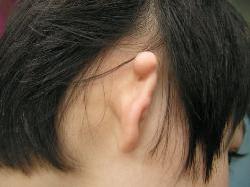
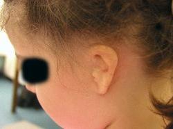
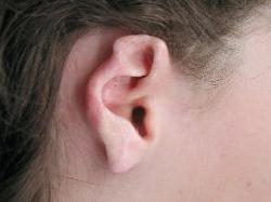
Ear Trauma Due To Piercing Ear Trauma Due To Burn |
|
The cause of Microtia is felt to be heterogeneous including genetic aberrations, teratogens (including isotretinoin, thalidomide, and maternal rubella), and vascular abnormalities. While no specific chromosomal abnormality has been cited for Microtia, amultifactorial inheritance is considered. Microtia is classified by Dr Firmin from Paris into type One, Two and Three. According to this classification we plan our surgical procedures. The evaluation for Microtia should include a thorough physical examination: evaluation of facial and ear structures, symmetry, and animation, and dental occlusal relationships. The quality of non-hair-bearing skin in the vicinity of the auricle, hairline, position of the remnant auricle, and future lobule, and condition of the contralateral ear must be evaluated. The patients should be referred to an otologist if middle ear or inner ear abnormalities exist. The ear with Microtia may have hearing problems associated with all aspects of the peripheral auditory mechanism eg: - Absent External Auditory Ear canal - Varying degrees of narrowing of the External Auditory Ear canal - Varying degrees of ossicle deformities from minor to complete absence - Absent middle ear cleft - Varying degrees of Cochlea (Hearing Organ) and Vestibular (Balance Organ) abnormalities including complete absence of the inner ear - Absence of the Auditory nerves (very rare) The chances of improvement in hearing depend on: - an accurate assessment of all of the above anomalies - the level and the type of hearing loss in the affected ear - the state of the opposite ear (is the unaffected ear normal) - the desire of the patient to undergo a difficult and prolonged surgical procedure Investigations the ear with Microtia include: - Tests of auditory function - CT scans of the Temporal bone (This must be performed in a centre where the Radiologist and Radiographer are skilled in performing the scan as well as interpreting the results) MRI scans to assess: - Cochlea - the Cochlea, Vestibular and Facial nerves Surgery: - Is difficult and prolonged - The incision is usually above the potential ear opening (Endaural Approach) and not behind the ear (Post Auricular approach). However sometimes the post Auricular approach is used if the ear canal reconstruction is being performed at the second stage of the Pinna reconstruction - At the Children's Hospital there are usually at least 2 operations performed under general Anaesthetic - At the first procedure the ear canal and Tympanic membrane is constructed - The second procedure occurs 2 weeks later to graft the newly formed ear canal - The ossicular reconstruction is usually not performed until it is certain that the External ear canal and the tympanic membrane is healed and stable Post Operative: - After the second grafting procedure there is regular visits to the Clinic for assessment of Hearing and for regular cleaning of the External Ear Canal - This will normally occur at about every 2-3 months for the first 2 years until the normal skin migration patterns are established Results: - In most patients the establishment of an ear canal occurs - Some 10-20% of patients require a revision of the ear canal - The hearing may become close to normal dependent on the pre-existing anatomy - The hearing is usually improved but not normal. Microtia Clinic At Westmead Children's Hospital Microtia is a condition in which one or both of the child's ears are found to be poorly developed at birth. This may vary from significant narrowing of the ear canal to a major deformity of both ear lobes to develop at all. In almost all cases this affects the child's self esteem, and because hearing is usually affected in one or both ears it can affect a child's learning and speech. The Microtia Clinic combines the services of plastics surgeon, an ear, nose and throat surgeon and paediatrician to assist children with Microtia. For some children, families just request a cosmetic surgeon to improve the appearance of the ear. For some children, improvement of hearing can obtained by surgery performed by an ENT specialist. The paediatrician assists by coordinating the clinic and by checking and advising on the language and educational progress of the child. |

|
About Microtia |


|
Copyright © 2008 John Vandervord, FRACS, All rights reserved. |